Gastric foreign bodies in a 5-year-old Exotic Shorthair Cat
- enquiries16342
- Oct 2, 2024
- 2 min read
Gastric foreign bodies in a 5-year-old Exotic Shorthair Cat
By Dr Christine Baker, Soundiagnosis September 2024
History:
A 5-year-old female spayed Exotic Shorthair cat was presented to a Melbourne veterinary clinic for vomiting, loose stools and weight loss. Physical exam revealed a palpable large firm abdominal mass. Abdominal ultrasound was recommended for further evaluation.
Soundiagnosis abdominal ultrasound findings:
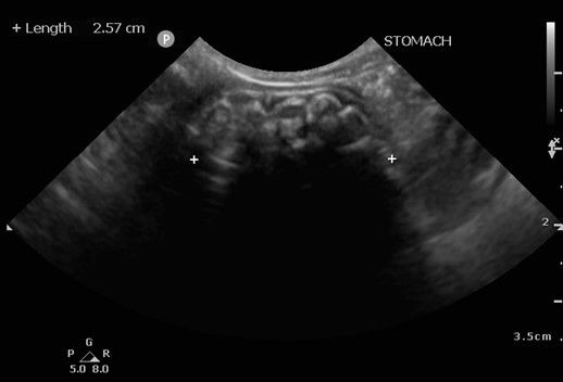
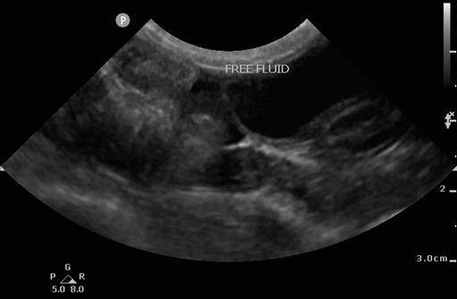
The ultrasound revealed severe dilation of the stomach with a large volume of heteroechoic content in the lumen which was casting strong distal acoustic shadowing. The content appeared to have irregular contours with bright hyperechoic areas within it. The remainder of the gastrointestinal tract displayed small amounts of anechoic fluid in the lumen but with no evidence of an obstructive pattern in the intestines. There was a small volume of anechoic abdominal effusion as well as diffusely hyperechoic peritoneal fat.
Diagnosis:
Gastric foreign material
Images:


Images 1 & 2: Marked dilation of the stomach with a large volume of heteroechoic content casting strong distal acoustic shadowing in the lumen.

Image 3: A small volume of anechoic abdominal effusion and diffusely hyperechoic peritoneal fat were also evident.
Comments and Outcome:
Foreign material in the gastric lumen was considered the most likely differential diagnosis for these imaging findings, and an exploratory laparotomy was recommended. A gastrotomy was performed and 16 elastic hair ties were successfully removed from the gastric lumen. The patient recovered well from surgery. This case highlights the variable appearance of gastrointestinal foreign bodies on ultrasound and the importance of imaging – the initial examination findings could have suggested a neoplastic mass in the abdomen but luckily for this patient the final diagnosis had a much better prognosis!


Comments