
Splenic mass in a 12 year old cat
- enquiries16342
- Sep 10, 2025
- 2 min read
By Dr Christine Baker, Soundiagnosis August 2025
History: The patient was presented for abdominal ultrasound due to a suspected abdominal mass and acute onset lethargy. She has recently had some hairballs but no regular vomiting. Blood tests revealed anaemia HCT 22%, neutrophilia, and mildly decreased platelets (118).
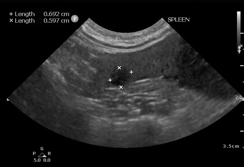
ULTRASOUND FINDINGS:
Hypoechoic irregular vascularised splenic mass. DDx. Neoplasia (lymphoma; mast cell tumour; sarcoma; other); nodular hyperplasia or extramedullary haematopoiesis less likely.
Cavitated hypoechoic non-vascularised tissue adjacent to the mass. DDx. Haematoma due to haemorrhage from the mass most likely; abscessation less likely.
Mild splenomegaly with one other well-defined hypoechoic nodule adjacent to the main mass, mildly mottled appearance of the parenchyma with a high frequency linear probe. DDx. Lymphoid hyperplasia; extramedullary haematopoiesis; emerging infiltrative neoplasia.
Diffusely hypoechoic abdominal lymph nodes preserving upper normal size. DDx. Reactive lymph nodes; emerging neoplasia.


OUTCOME: A splenectomy was performed and the splenic mass was sent for histopathology. The mass was diagnosed as a splenic stromal sarcoma (mitotic count 16) most likely arising from complex nodular hyperplasia.
COMMENTS: Splenic stromal sarcomas are rare in cats, but limited information in dogs suggests 1 year survival rate of 90% after complete resection. In general, large splenic masses are less common in cats than dogs. Mast cell neoplasia and lymphoma are the most common types of splenic neoplasia seen in cats and may cause diffuse splenomegaly or masses or nodules in the spleen. Haemangiosarcoma can also occur in cats. As these conditions can involve other abdominal organs, complete abdominal ultrasound is important in cats with splenic masses or splenomegaly.


Comments